



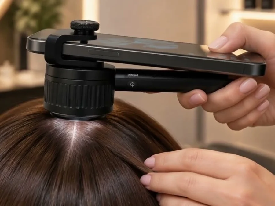
In short: trichoscopy is a non-invasive, magnified examination of the scalp and hair that is painless, quick and repeatable; it is the first-line tool for most hair loss. A scalp biopsy removes a small sample of skin under local anesthetic for histopathology and is reserved for suspected scarring alopecia, unclear cases or when disease activity must be confirmed. Most people with common hair loss do not need one.
Both exams try to answer the same question — what is happening to the follicles — but at different depths and with very different levels of invasiveness. This guide compares trichoscopy vs biopsy, explains what each can and cannot show, and clarifies when a dermatologist is likely to recommend a scalp biopsy for hair loss.
What trichoscopy shows — and what it cannot
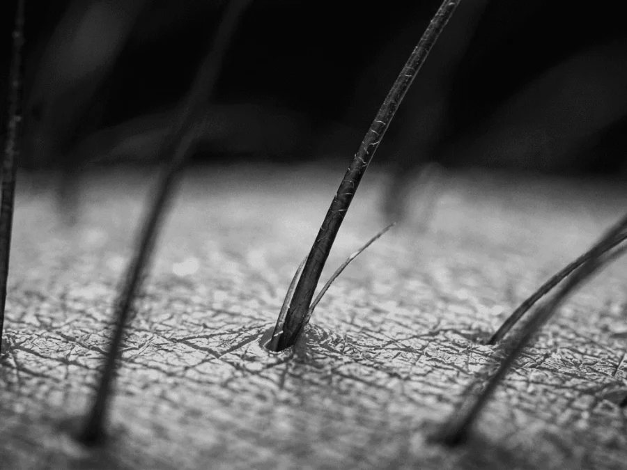
Trichoscopy is dermoscopy applied to the scalp: a handheld or video dermatoscope magnifies the skin surface and the hair shafts to reveal details that are invisible to the naked eye. Nothing is cut, no anesthetic is needed, the exam takes minutes, and it can be repeated at every visit to document how a condition evolves — one of its greatest strengths. If you are new to the method, our beginner chapter on the trichogram and trichoscopy explains the basics.
What the examiner is looking for:
- Hair shaft diameter variability — thin and thick hairs side by side, a typical feature of pattern hair loss linked to androgen sensitivity.
- Follicular openings — whether they are preserved or lost; loss of openings raises the suspicion of a scarring alopecia.
- Yellow dots, black dots, exclamation-mark hairs and broken hairs, which help separate alopecia areata, trichotillomania and tinea capitis.
- Perifollicular scaling and redness, and the pattern of visible vessels, which point to inflammation around the follicle.
Trichoscopy is useful for androgenetic alopecia, alopecia areata, telogen effluvium (largely a diagnosis by exclusion once other patterns are ruled out), clues to tinea capitis, trichotillomania, and for suspecting scarring alopecia. Its limits matter just as much: it sees only the surface and cannot look inside the follicle or the dermis, some findings overlap between conditions, and the result depends on the operator’s experience and the device used.
What a scalp biopsy involves
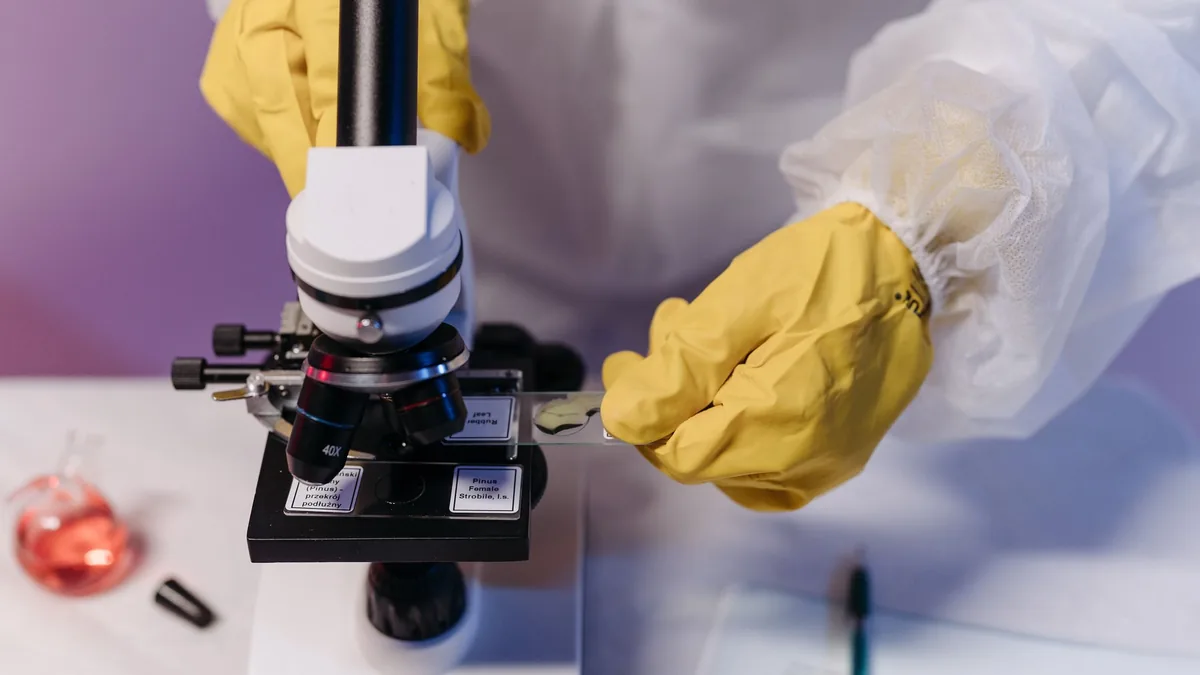
A scalp biopsy removes a small cylinder of skin — usually a 4 mm punch biopsy — under local anesthetic. The dermatologist typically takes one or two samples, sometimes closing the site with a stitch or two. The tissue goes to a dermatopathologist, who examines it under the microscope in horizontal (transverse) and/or vertical sections: horizontal sections allow many follicles to be assessed at once, while vertical sections show a follicle through its full depth together with the surrounding skin.
Results typically take several days to a couple of weeks. The procedure leaves a small scar, usually hidden by the surrounding hair, and carries the minor risks of any small skin procedure: bleeding, infection, pain and scarring. In exchange, the biopsy answers a question trichoscopy cannot: what is actually happening in and around the follicle, below the surface — inflammation, fibrosis, miniaturization, or a process that is not a hair disorder at all.
Trichoscopy vs biopsy: a side-by-side comparison
- Invasiveness: trichoscopy is non-invasive; a biopsy removes a small piece of skin.
- Anesthetic: none for trichoscopy; local anesthetic for a biopsy.
- Time: trichoscopy takes minutes and gives an immediate impression; biopsy results take days to a couple of weeks.
- What it sees: trichoscopy shows the surface — shafts, openings, scaling, vessels; histopathology shows the follicle and dermis in depth.
- Repeatability: trichoscopy can be repeated at every follow-up; a biopsy is done once or twice in most cases.
- Risks: essentially none for trichoscopy; a small risk of bleeding, infection, pain and a small scar with a biopsy.
- Typical role: trichoscopy for initial diagnosis and monitoring of most hair loss; biopsy for suspected scarring alopecia, unclear cases and confirming disease activity.
The two are complementary rather than competing. In practice, a biopsy is almost always preceded by a clinical examination and trichoscopy, and the findings of one are interpreted in light of the other. Our article on what trichoscopy is and how a hair analysis works covers that first step in more detail.
When a scalp biopsy for hair loss is usually considered
Most patients with common hair loss — pattern hair loss or telogen effluvium — are diagnosed from the history, the clinical exam and trichoscopy and never need a biopsy. A dermatologist usually considers one in the following situations:
- Suspected scarring (cicatricial) alopecia such as lichen planopilaris, frontal fibrosing alopecia, discoid lupus erythematosus, folliculitis decalvans or central centrifugal cicatricial alopecia, where the diagnosis has treatment implications and follicle loss is permanent.
- An unclear diagnosis after clinical examination and trichoscopy.
- Distinguishing diffuse alopecia areata from telogen effluvium or androgenetic alopecia when the surface findings overlap.
- Confirming disease activity in a known scarring alopecia, to guide how actively it needs to be treated.
- Suspicion of another scalp disease, including tumors, that presents with hair loss or a changed patch of skin.
Scarring alopecia: why the diagnosis should not wait
In non-scarring hair loss the follicle survives, even if it shrinks or rests for too long. In scarring alopecia the follicle is replaced by fibrous tissue, and once that has happened the hair in that spot does not return. This is why the decision to biopsy carries a sense of urgency: the aim is to recognize the process while it is still active and the remaining follicles can still be protected. Our beginner guide to hair loss and alopecia describes the main scarring types.
Signs that justify seeing a dermatologist promptly rather than waiting to see how things develop:
- Itching, burning or pain in the scalp that accompanies the hair loss.
- Redness around the follicles, scaling that hugs the base of the hairs, or pustules.
- A smooth, shiny patch of scalp in which the follicle openings are no longer visible.
- A receding frontal hairline, often band-like, together with thinning or loss of the eyebrows.
Trichoscopy-guided biopsy: choosing the right site
One reason the two exams work so well together is that trichoscopy helps the dermatologist decide where to take the sample. A biopsy from a burnt-out, end-stage area may show little more than scar tissue, which confirms that follicles have been lost but says nothing about the cause. Trichoscopy makes it possible to identify the active border of a lesion — where perifollicular redness, scaling or inflammation is still visible — and to place the punch exactly there. This improves the diagnostic yield of the sample and, in many cases, may spare the patient a second procedure.
Preparing for a scalp biopsy
A punch biopsy is a short outpatient procedure, but a little preparation helps it go smoothly and reduces the small risks involved:
- Tell the dermatologist about blood thinners — anticoagulants and antiplatelet medicines — and about any bleeding tendency.
- Mention allergies, especially to local anesthetics, antiseptics or dressings.
- Do not stop any medication on your own; if an adjustment is needed, the doctor will tell you how and when.
- Ask for the aftercare instructions: how to keep the site clean and dry, when to wash your hair, how to recognize signs of infection, and when any stitches will be removed.
Mild tenderness for a day or two is expected. Increasing pain, spreading redness, pus, or bleeding that does not stop with gentle pressure should be reported to the clinic.
Who decides — and what an online diagnosis can do
The decision to take a scalp biopsy belongs to a dermatologist, who weighs the clinical picture, the trichoscopy findings and the likely benefit of a histological answer. Trichology specialists working from photographs and a questionnaire can do something different but valuable: triage. From good images of the scalp and a clear account of the symptoms, they can often tell whether a pattern looks like common hair loss that may be monitored with routine follow-up, or whether there are warning signs that call for an in-person examination and, possibly, a biopsy.
What online diagnosis cannot do is replace the biopsy itself, or the dermatologist who performs it. If you are unsure where your own hair loss sits, a free assessment through our online Hair Clinic can be a reasonable first step: a trichology specialist reviews your photos and questionnaire and indicates whether a face-to-face examination is advisable, with no obligation.
Frequently asked questions
No. Trichoscopy is a magnified look at the scalp and hair with a dermatoscope. Nothing is cut or pulled, no anesthetic is used, it takes a few minutes, and it can be repeated as often as needed to follow a condition over time. In most cases, no. Pattern hair loss and telogen effluvium are usually diagnosed from the history, the clinical exam and trichoscopy. A biopsy is considered when a scarring alopecia is suspected, when the diagnosis remains unclear, when diffuse alopecia areata must be distinguished from other causes, or when another scalp disease is suspected. A 4 mm punch biopsy leaves a small scar, usually hidden by the surrounding hair. Minor risks include bleeding, infection and some pain. Following the aftercare instructions helps keep the scar as small as possible. Not really — they look at different things. Trichoscopy sees the surface and is ideal for diagnosis and follow-up of most hair loss; a biopsy looks inside the follicle and dermis. Trichoscopy also helps choose the most informative site when a biopsy is needed, and an online assessment can triage but never substitute for it.Is trichoscopy painful or harmful to the hair?
Do I need a scalp biopsy for hair loss?
Will a scalp biopsy leave a scar?
Trichoscopy vs biopsy: can one replace the other?
This article is part of our guide to trichogram and trichoscopy. You may also find these useful: preparing for a trichoscopy and reading hair analysis results.
This article is for general information and is not a substitute for personalized medical advice. SIMONE TRICHOLOGY products are dermocosmetic and are not intended to diagnose, treat, cure or prevent any medical condition.