

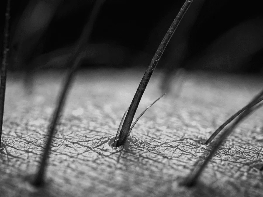


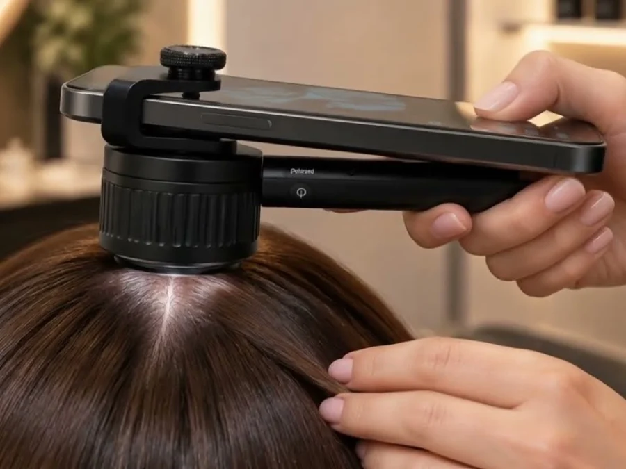
In short: hair analysis results from trichoscopy, a trichogram or a digital scalp analysis describe what hair and scalp look like under magnification: density, hairs per follicular unit, shaft diameter and its variability, miniaturized hairs, the growing-to-resting ratio and scalp signs such as scaling or redness. Reference values vary by device and area, one report is only a snapshot, and the diagnosis is made by a professional.
If you have just received a scalp analysis report full of numbers, percentages and close-up images, this guide walks you through hair analysis interpretation step by step: what each metric measures, which findings deserve attention, what the report cannot tell you, and the questions worth asking your specialist.

First, which hair analysis are we talking about?
In trichology, “hair analysis” means examining the hair and scalp under magnification: trichoscopy (a dermatoscope or digital camera on the scalp), a trichogram (a small sample of plucked hairs examined under a microscope) or a phototrichogram, in which the same area is photographed a few days apart to see how many hairs are growing. If these methods are new to you, start with our hub chapter on trichogram and trichoscopy and our explainer on what trichoscopy is and how it is performed.
It is not the same as a “hair mineral analysis” or hair toxicology test, which measures substances inside cut hair shafts. Those tests are sometimes marketed as a way to detect nutritional deficiencies, but this use is not supported by good evidence: mineral hair tests are not a reliable way to diagnose a deficiency, and doctors use blood tests for that. Everything below refers to image-based analysis.
One more thing before reading any figure: reference ranges in a scalp analysis report depend on the device and its software, the method, the exact area measured, your age, ethnicity, and your hair color and type. A fine blond scalp and a coarse dark one can be equally healthy and produce very different readings. Compare your results with the references your clinic or device provides, not with numbers found online; the sections below deliberately avoid quoting “normal” values.
Hair density and follicular units
Hair density is expressed as hairs per square centimeter (hairs/cm²) in the area captured. Many reports also give follicular unit density, the number of follicular groupings in that same square centimeter, and from the two they calculate hairs per follicular unit.
- Hairs per follicular unit: units carrying two or three hairs are the usual picture on a healthy scalp. A high proportion of single-hair units can suggest miniaturization, the gradual shrinking of follicles seen in androgen-sensitive hair loss.
- Density by area: a report that measures the frontal area or crown as well as the back of the head (the occipital area) is more informative than a single spot, because the occipital area is often used as the internal reference against which other areas are compared.
- Empty follicles: follicular openings with no visible hair may appear during active shedding or where follicles have stopped producing a visible shaft.
Hair shaft diameter, variability and miniaturized hairs
Shaft diameter is the thickness of individual hairs, usually stated as an average plus a measure of variability (how much the hairs in one area differ from each other). Two readings deserve particular attention in hair analysis interpretation:
- Diameter variability: in androgenetic alopecia, hairs in one area tend to differ more from each other, with a growing number of thin hairs, especially in the frontal and crown areas compared with the occipital reference.
- Proportion of vellus or miniaturized hairs: these are thin, short, poorly pigmented hairs. A rising share of them in the areas prone to androgen sensitivity is one of the typical signs the specialist looks for.
A low average diameter on its own is not alarming if it is uniform across the scalp and matches your hair type. It is the pattern, the comparison between areas and the change over time that carry meaning.
Trichogram results: anagen, telogen and dystrophic hairs
A trichogram or phototrichogram reports the proportion of hairs in each phase of the growth cycle. Anagen hairs are growing; telogen hairs are resting and about to be shed. On a healthy scalp, the large majority of hairs are in anagen at any given time, and hair grows at roughly 1 cm per month during this phase.
- A higher-than-expected share of telogen hairs suggests increased shedding, as in telogen effluvium, which often follows a trigger (illness, childbirth, a demanding period, a change in medication) by a few months.
- Dystrophic hairs have abnormally shaped roots and can appear when growth has been abruptly disturbed.
- Broken hairs usually point to fragility of the shaft, although they can also be seen in some scalp conditions.
Keep in mind that trichogram results are sensitive to conditions: when you last washed, how the sample was taken and from which area all influence the percentages, which is one more reason to interpret them with the professional who performed the test.
Scalp findings in your report
Alongside the hair metrics, a scalp analysis report describes the skin itself. Some findings are mainly dermocosmetic; others are signals that a medical assessment is needed.
- Sebum and oiliness: a shiny film, greasy residue or clogged openings point to an oily scalp pattern.
- Scaling or flaking: fine, loose scales often go with a dry pattern; thicker, greasy, adherent scales with an oily one.
- Redness (erythema) and vessel patterns: diffuse redness or distinctive blood-vessel arrangements can reflect irritation or inflammation.
- Perifollicular scaling or redness: when concentrated right around the follicle openings, this can indicate inflammation and deserves attention.
- Yellow dots, black dots and exclamation-mark hairs: these trichoscopic signs are associated with specific conditions, such as alopecia areata, and help the specialist narrow down the picture.
- Loss of follicular openings: smooth areas where the openings can no longer be seen are a warning sign for scarring alopecia and should be reviewed by a dermatologist without delay, because in scarring processes the follicle may be permanently lost.
Hair shaft condition: cuticle, split ends and breakage
Magnified images of the lengths show the state of the cuticle (lifted or eroded scales), split ends and points of breakage. These are usually cosmetic findings linked to heat, chemical processing, friction and brushing, and they respond to changes in hair care rather than to scalp treatment. They matter because breakage along the length is often mistaken for hair loss from the root.
Scores, snapshots and trends over time
Many digital devices and apps summarize the capture in an overall score or index. These are proprietary: each manufacturer decides what goes into the number and how it is weighted, so a score from one system cannot be compared with another. Ask what the score includes and which individual metrics drive it. Our article on AI scalp analysis and digital trichology explains what an automated capture actually measures.
Whatever the format, remember three principles:
- One result is a snapshot. Density and telogen ratio fluctuate with season, stress, illness and the moment in your hair cycle.
- Trends matter more than single values. The useful question is whether frontal density, the miniaturization share or the telogen percentage is moving over months.
- Comparisons must be like for like: same area of the scalp, same lighting and magnification, and the same timing after washing. Otherwise the “change” may simply be a change in conditions.
What the report cannot tell you
An analysis describes signs; it does not explain them. It can show more telogen hairs, but not whether the cause is a thyroid issue, low iron stores, a medication or a recent illness. It can show miniaturization in the pattern typical of androgen sensitivity, but not your hormone levels. It cannot measure vitamins, minerals or inflammation markers. For those questions, a doctor combines your history with blood tests or, when a scarring process is suspected, a scalp biopsy (typically a 4 mm punch).
The diagnosis is therefore made by a professional who puts the images together with your history, family background, medications and, when needed, laboratory results. Questions worth asking at your appointment include:
- Which areas were measured, and is there an occipital reference for comparison?
- Which findings are most relevant in my case, and which are within the expected range for my hair type?
- Are there signs of inflammation or loss of follicular openings that need a dermatologist?
- Do you recommend blood tests to look for a cause?
- When should the analysis be repeated, and under which conditions, so the results are comparable?
Next steps after your hair analysis results
Because the hair cycle is slow, changes take time to become visible, and a follow-up analysis is often scheduled every few months rather than every few weeks. Keep your reports and images together, note the date and the time since your last wash, and bring any blood test results to the next review. If your report mentions inflammation, scarring signs or a rapid change you cannot explain, see a dermatologist. If you would like a guided first reading of your hair and scalp by a trichology specialist, the free online Hair Clinic assessment from photos and a short questionnaire is a simple, no-obligation way to start.
Frequently asked questions
There is no single figure. Density in hairs per cm² varies with ethnicity, age, hair color, hair type, the area measured and the device used. Compare your results with the references of the clinic or device that produced them, and pay more attention to the trend over time and to differences between areas of your scalp than to one absolute number. A higher share of resting hairs usually means more hairs are being shed than expected, as in telogen effluvium, which often follows an illness, childbirth, a stressful period or a medication change by a few months. The trichogram shows the shedding; the cause is identified by a professional, sometimes with blood tests. No. Trichoscopy and trichograms describe the hair and scalp; they do not measure iron, vitamins or hormones. Hair mineral tests are not a reliable way to diagnose deficiencies either. A doctor uses blood tests for those questions and combines them with the images and your history. Often every few months, because hair cycles are slow and earlier comparisons may reflect normal fluctuation rather than real change. Repeat it in the same area, with the same lighting and the same timing after washing, so that the results are comparable.What is a good hair density in hair analysis results?
What do trichogram results with a high telogen percentage mean?
Can a scalp analysis report diagnose iron deficiency or a hormonal problem?
How often should I repeat the hair and scalp analysis?
This article is part of our guide to trichogram and trichoscopy. You may also find these useful: preparing for a trichoscopy and trichoscopy vs scalp biopsy.
This article is for general information and is not a substitute for personalized medical advice. SIMONE TRICHOLOGY products are dermocosmetic and are not intended to diagnose, treat, cure or prevent any medical condition.